MR Linac | Magnetic resonance-guided linear accelerator for radiation therapy
Having the MR Linac available to treat oncology patients is a great therapeutic advantage in the administration of radiotherapy.
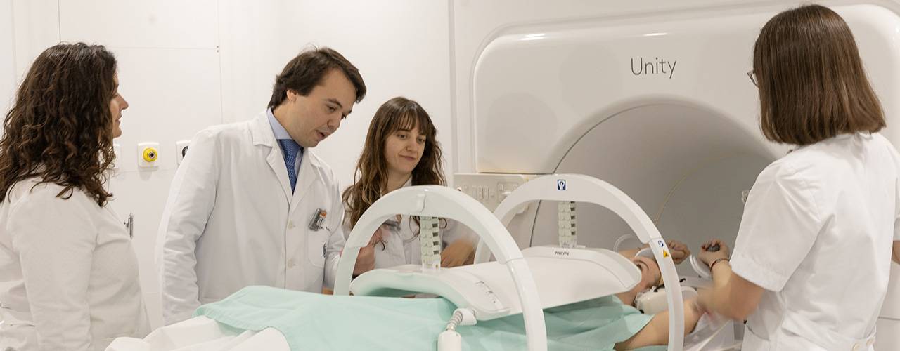
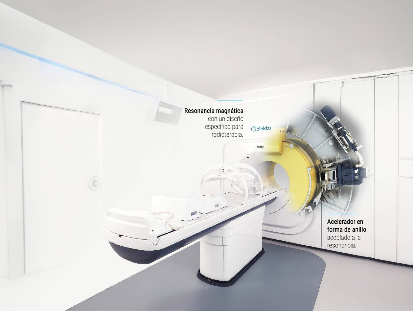
MR Linac is an advanced radiotherapy technology that combines a linear accelerator with an integrated 1.5 Tesla magnetic resonance imaging system, with high diagnostic accuracy before, during and after treatment.
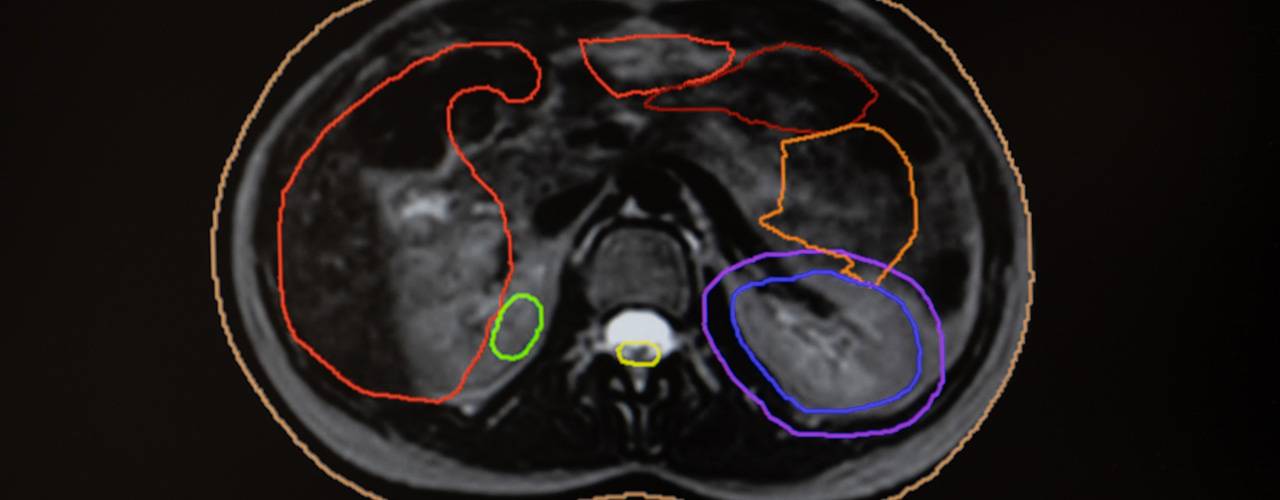
At present, in most hospitals, radiotherapy treatment planning is carried out using CT (computed tomography) images, but this is not very accurate in visualising soft tissue and other areas, especially in the abdominal area. However, with the help of the MR Linac's 1.5 Tesla MRI, these tissues can be visualised with high clarity and definition.
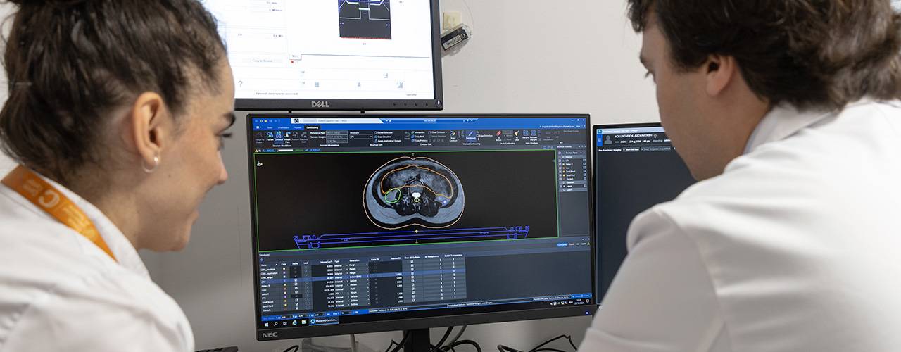
During the sessions and over the duration of the treatment, the tumour changes in shape and position. MR Linac technology makes it possible to administer adaptive radiotherapy in real time, which adapts the dose and precision of the radiotherapy administered to the patient according to the specific characteristics of each person and each tumour.
With the acquisition of this equipment at the Pamplona site, the Clínica Universidad de Navarra has become the best technologically equipped Radiation Oncology department in Spain and one of the most advanced in Europe.

Advantages of the MR Linac accelerator
Real-time images
Integrated magnetic resonance imaging allows visualization of internal body movements, adjusting the dose or target of radiotherapy instantly.
Improved accuracy
It allows increasing the dose of radiotherapy administered only to the tumor tissue, which will increase efficacy and reduce the number of sessions required.
Protection of healthy tissues
High-quality images obtained in real time during treatment delivery minimize damage to surrounding healthy tissues.
Adaptive radiation therapy
The team of professionals are present during the session to assess the changes that occur in real time and adjust the dose to maximize its effectiveness.
How does the resonance-guided accelerator work?
MR Linac allows physicians to observe your tumor as they treat it, and adapt the dose of radiation therapy to changes in the tumor in real time.
Which tumors can be treated with MR Linac?
A customized program for each tumor type and patient characteristics

Pancreatic Cancer
One of the most difficult types of tumors to treat due to its location and proximity to vital organs is pancreatic cancer, which benefits significantly from MR Linac. The ability to adjust the treatment live as the tumor oscillates to the rhythm of internal body movements, such as breathing, allows for safer and more effective radiotherapy, minimizing the risk of damaging adjacent structures.

Rectal Cancer
The high precision of this technology makes it possible to minimize damage to healthy tissues surrounding the tumor, such as the bowel or pelvic organs. This makes it possible to increase the dose of radiotherapy applied to the tumor to avoid surgery, the consequences of which significantly affect patients' quality of life.

Prostate Cancer
Prostate tumors, often treated with radiotherapy, also find great advantages with MR Linac. This technology allows immediate adjustments in radiotherapy delivery, even in case of changes in the patient's anatomy, thus improving treatment accuracy and shortening the number of sessions required.

Bladder Cancer
Combining radiotherapy applied with MR Linac, together with the administration of chemotherapy, makes it possible in many cases to avoid bladder removal (cystectomy). This precision radiotherapy improves the effectiveness of treatment and reduces side effects in patients with bladder cancer.

Brain Tumors
In the treatment of brain tumors, the MR Linac is especially useful thanks to its millimeter accuracy. Brain tumors are often close to critical areas of the brain and accuracy is essential in their approach. The integrated MRI allows specialists to monitor anatomical movements during the radiation therapy session, thus ensuring that the radiation is targeted exactly and exclusively at the tumor.

Metastases
This technology especially benefits the treatment of metastases affecting moving organs or sensitive structures, such as the lungs, liver and brain. In these cases, it is an alternative to surgery or other local treatments, such as radiofrequency or embolization, reducing the side effects and sequelae that can appear after these procedures.
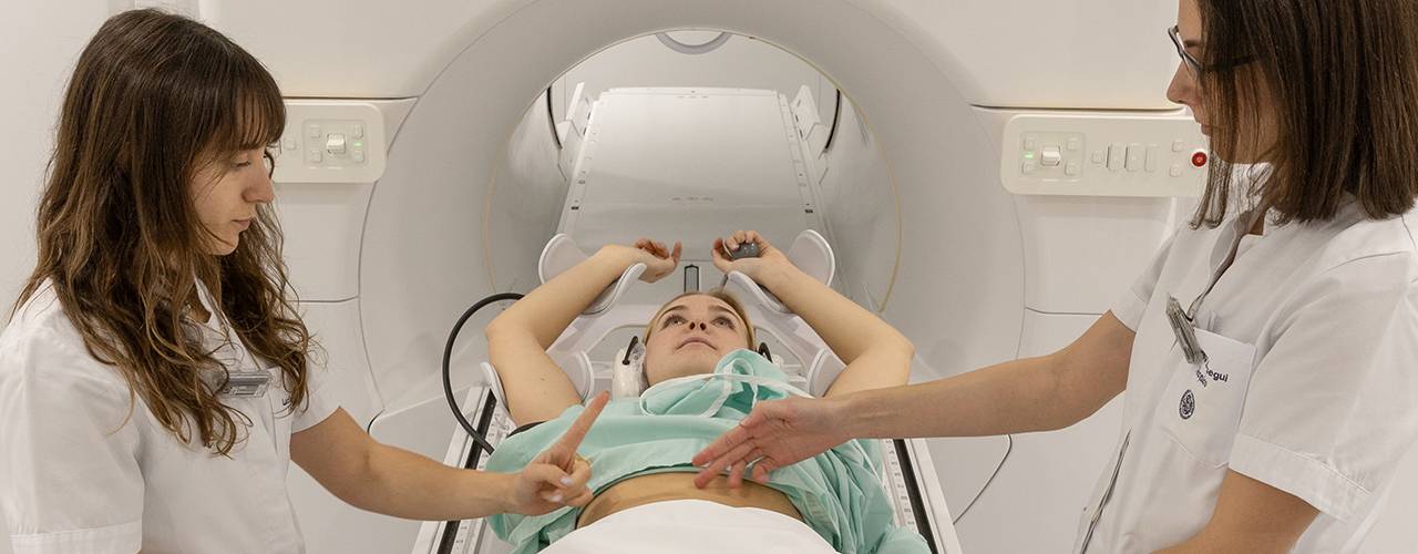
How is the patient's process during treatment?
Research
Our lines of research will be aimed at personalizing the treatments we offer to our patients and understanding the response of tumors with adaptive radiotherapy.
- Functional imaging: analysis of MRI image sequences to assess tumor response to treatment on a daily basis.
- Radiomics: quantifiable imaging parameters that correlate with prognostic and response patterns of different tumors.
- Dosimetry: precise calculation of the radiation dose received each day to redefine the safety limits for healthy tissues.
- Clinical programs: prospective research projects that will help collect data during treatment to evaluate its effectiveness and future approaches.
Our team of professionals
Radiotherapy Oncology Specialists
Professionals from the Radiophysics and Radiation Protection Service


