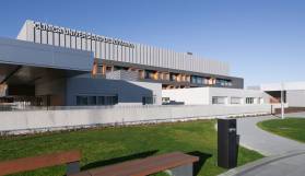
External Radiation Therapy
"We incorporate systems that make it possible to administer curative doses in the risk area and minimize radiation to healthy adjacent tissues, according to the characteristics of each patient".
DR. MARTA MORENO SPECIALIST. RADIOTHERAPEUTIC ONCOLOGY DEPARTMENT

What is external radiation therapy?
In external radiotherapy, ionizing radiation beams (electromagnetic waves) generated in radiation equipment far away and external to the patient (linear accelerators) are used.
The Radiation Oncology Department has the VERSA HD accelerator, considered the most advanced for external radiotherapy treatments. It offers a treatment speed up to ten times higher than that of any conventional accelerator, together with greater precision in the administration of the dose.
In the external radiotherapy program, stereotactic techniques and intensity-modulated radiotherapy are available systematically for most tumors. The treatment techniques are the conventional ones - external radiotherapy and three-dimensional conformal external radiotherapy - and also special ones - 3D external radiation, modulated external radiotherapy and, as mentioned above, stereotactic radiotherapy (radiosurgery and extracerebral stereotactic radiotherapy).

A PERSONALIZED MEDICINE
Second Opinion,
peace of mind
Request a second opinion from our professionals with great experience in the diagnosis and treatment of oncological diseases
In 3 days, without leaving home.
MR Linac | MRI-guided linear accelerator for radiation therapy
Advanced radiotherapy technology that combines a linear accelerator with an integrated magnetic resonance of 1.5 Teslas, it allows adaptive radiotherapy to be administered in real time, which adapts the dose and precision of the radiotherapy administered to the patient according to the characteristics of each person and each tumor.
Types of external radiotherapy
The Clinic has extensive experience in three-dimensional conformal external radiotherapy (RT3D). This technique incorporates CT technology into the process of designing radiation treatments.
Another advance incorporated is the development of three-dimensional (3D) mode treatment planners, based on complex computerized calculation systems to estimate radiation dose distribution.
All this implies quality, since with the RT3D the spatial distribution of the radiation is improved, adapting better to the volume and shape of the tumor. It can significantly decrease the amount of radiation on the adjacent healthy organs and the treatment can be administered to cure without resorting to surgery.
The equipment undergoes daily quality control. We have three state-of-the-art linear accelerators and the latest and most advanced planning systems.
The shape of the radiation treatment field in conventional RT3D is made by a manual process almost handmade. Blocks are made of a manually carved leaded material which will be given shape and size according to the tissue or organs to be protected.
Elaborated and carved, the blocks are assembled in trays that are interposed to the exit of the radiation beam. This interposition casts a shadow over the field corresponding to the area or tissues to be protected.
Intensity-Modulated Radiation Therapy (IMRT), the latest generation of external radiation treatments
It is only available at highly specialized oncology treatment centers.
The Clinica Universidad de Navara is one of the pioneering Spanish institutions in the use of this special radiation technique, with experience in the treatment of patients of more than 15 years.
This treatment allows the shaping of the treatment fields and modulates the intensity of the radiation beams by means of multileaf or multileaf collimators controlled from the planner in a completely digital environment. With this technique, each of the multiple fields provided is subdivided by the collimator into multiple segments.
This feature and a series of other sophisticated improvements, such as inverse planning (drawing up treatment plans based on proposals for dose levels and restrictions for healthy target tissues and organs) make it possible to draw up highly conformed treatment plans with differentiated radiation dose distribution maps within the same field.
This technique makes it possible to apply more intelligent treatments with a potential for increased efficacy derived from the possibility of scaling radiation doses with a higher safety profile.
Stereotactic radiotherapy is a special external radiation technique.
It requires special fixation and immobilization systems (stereotactic guides or frames) with a three-dimensional coordinate system, independent of the patient, which allow the lesions to be located with high precision.
At the same time, it requires radiation therapy equipment that generates highly conformed radiation beams (linear accelerators, gammaknife, cyberknife, tomotherapy) that converge on the lesion by ultraselectively administering high doses of radiation on it without increasing the radiation on adjacent healthy organs or structures.
Several treatment sessions can be applied with very high precision. The most frequent use is the treatment of brain tumors.
SBRT (Stereotactic Body Radiation Therapy) represents the radiation under stereotaxic conditions of extracerebral lesions. It is possible to treat lesions, such as inoperable lung tumors or liver metastases, in a short time and with a tolerable side effect profile.
Applying a single treatment session with very high doses of radiation administered through stereotaxic systems and conditions is radiosurgery.
It is indicated in the treatment of malignant or benign lesions smaller than 3-4 centimeters (brain metastases, arteriovenous malformations, neurinomas, meningiomas).
The Clínica Universidad de Navarra is the first private hospital in Spain that has acquired a miniaturized linear accelerator to administer intraoperative radiotherapy: LIAC, the most versatile model, with a smaller size and greater mobility.
The LIAC makes it possible to treat more complex patients by reducing to a minimum the level of toxicity in healthy adjacent tissues, in fewer sessions and in a safer manner.
The time of exposure to radiation does not exceed two minutes, since the accelerator makes it possible to achieve a very high and homogeneous dose in a very specific area in a single session; a single session of this accelerator is equivalent to six sessions of conventional radiotherapy.
(only in spanish)
Do you need to request a consultation with one of our specialists?
How is external radiotherapy performed?
A specially trained team of professionals led by a radiation oncologist participates in the radiotherapy treatment.
The radiation oncologist is a physician specializing in radiation oncology who develops, prescribes and supervises the radiation treatment plan. He can modify the treatment depending on the patient's evolution, identifies and treats the adverse effects of irradiation and collaborates with other specialists involved in the multidisciplinary treatment of cancer such as medical oncologists and surgeons.
Medical physicists work closely with the radiation oncologist in planning and administering treatment. They supervise the work of the dosimetrist and are directly involved in planning complex treatments. In addition, they develop and manage treatment unit quality programs and perform tests to establish the proper functioning of the units and the quality of the radiation beam.
The dosimetrists work together with the radiation oncologist and the medical physicist to select the radiation technique capable of generating the best distribution of the radiation dose over the tumor and the greatest exclusion of radiation doses to healthy tissues. The work is done on computers that use complex calculation algorithms capable of processing different types of images.
The radiotherapy technician is the person in charge of performing the daily radiation treatment supervised by the physician. He or she must be meticulous in the daily immobilization and positioning of the patient, ensure that the proper treatment has been done, and make a daily record of the treatment.
The radiation oncology nurse works with the entire treatment team to address the needs of the patient and family before, during and after treatment. They explain the care to be taken during and after irradiation and possible adverse effects and how to treat them.
Other health professionals involved in the care of these patients include medical nutritionists, physical therapists, dentists, and social workers.
Prior to treatment with radiation therapy, the medical radiation oncologist talks with the patient and explains the benefits and risks of treatment as well as other existing therapeutic possibilities.
Afterwards, the simulation is performed, which consists of taking measurements and drawing references on the skin to facilitate the entry of external radiation beams through the skin in a precise and reproducible way in each of the treatment sessions. The patient is immobilized in a comfortable and reproducible position that will be used daily during the irradiation.
For the immobilization of the patient, different devices are used such as thermoplastic masks, vacuum or catalytic resin mattresses, inclined planes, etc., selecting a certain method of immobilization depending on the tumor location and the required precision of the case.
Under these conditions of immobilization and fixation of the patient, a planning CT scan is performed and the corresponding axial images are acquired. These CT images are sent to a computer for virtual planning of the radiation treatment. In the planning computer, a certain photon energy, the number of radiation fields (usually two to four) and the rotation angles of the accelerator head are chosen.
Several treatment plans are generated and the radiation oncologist selects the plan that presents an optimal radiation dose distribution capable of maximizing the radiation dose to the tumor while minimizing the dose to adjacent normal structures.
Finally, the patient starts the treatment in the same position in which the simulation and planning procedures have been performed, after verifying the radiotherapy fields, which is done by comparing images reconstructed in the virtual planning with real images of the patient himself generated by means of a radiographic plate or digital portal images.
The treatment with radiotherapy is painless and the patient does not notice any type of sensation. It is administered on an outpatient basis in the form of daily sessions (fractions), five days a week, from Monday to Friday, resting on Saturdays and Sundays up to a total of 25-40 fractions depending on the type of tumor treated.
Sometimes the treatment has to be interrupted for a day or more due to the appearance of adverse effects and is resumed when these have improved or disappeared.
During the treatment, check-up X-rays and weekly check-ups are scheduled with the radiotherapy oncologist and the nurse to observe the evolution, the adverse effects and recommend treatment if necessary.
It is usual to recommend to patients certain care and habits during and after the irradiation such as getting enough rest, following a balanced and nutritious diet and paying special attention to the skin that may become sensitive and red.
After treatment, the patient is scheduled for an appointment to check on his recovery and the evolution of the cancer.
In general, radiotherapy is well tolerated and many patients are able to carry out their normal activity, however, in some patients adverse effects may appear that are generally limited to the treated area.
The adverse effects of radiotherapy are acute when they occur during the treatment period and in the ninety days that follow. They are the result of an inflammatory process derived from the depletion of progenitor cells from rapidly growing tissues such as skin, mucous membrane of the oral cavity and gastrointestinal tract, hematopoietic tissue, hair follicle, etc.
These acute adverse effects are transitory and recoverable due to the repair capacity of healthy tissue. They usually appear after the second or third week of treatment and may last for several weeks after treatment.
The most frequent symptom is fatigue, which is not usually disabling. Mucositis (inflammation of the oral mucosa), esophagitis (inflammation of the esophagus), enteritis (inflammation of the small intestine), epithelitis and dermatitis (inflammation of the skin), alopecia and spinal cord aplasia are the most frequently observed acute adverse effects. On many occasions it is necessary to administer anti-inflammatory treatment, feeding with special means and less commonly, hospital admission for hydroelectrolyte replacement.
Chronic adverse effects are observed 90 days after the end of radiotherapy and are the result of a process of tissue transformation derived from the depletion of slow-growing cells such as muscle, renal and hepatic parenchyma, nervous tissue, etc.
These chronic adverse effects are not recoverable and are permanent, constituting the most important limiting factor of clinical radiotherapy, although it is true that the probability of the appearance of chronic adverse effects is low.
Among the chronic adverse effects are xerostomia or loss of saliva, fibrosis or hardening of the subcutaneous tissue, lung or intestine, necrosis, neurological damage and in the pediatric population, growth retardation, hormonal alterations and the appearance of second tumors.
The Department of Radiation Oncology of the Clínica
of the Clínica Universidad de Navarra
The Clínica Universidad de Navarra's Department of Radiation Oncology has extensive experience in external and intensity-modulated radiotherapy. In addition, we apply various state-of-the-art medical-surgical techniques available in a few Spanish centers.
We are one of the international reference centers in the performance of intraoperative implants and radiation treatment with high rate brachytherapy technique during the postoperative period.
We have one of the most extensive experiences worldwide in the treatment of intraoperative brachytherapy of head and neck tumors, soft tissue sarcomas and gynecological tumors.
Treatments we perform

Why at the Clinica?
- Expert professionals of reference at international level.
- Greater accessibility for national and international patients.
- State-of-the-art technology, the most advanced in Spain.
- The most advanced proton therapy unit in Europe at the Madrid headquarters for the treatment of cancer with protons.


